CASE-
A 55 year old female came to the opd with the chief complaints of
1.palpitations (since 2months)
2.chest pain (since 2 months)
3.associated with shortness of breath
HOPI-
patient was apparently asympotamatic 2months back then she started having chest pain on left side more on epigastrium, non-radiating
-associated with palpitations
-and started developing shortness of breath, around the same time, progressive in nature leading to present status of grade III-IV of dyspnoea.
-associated with PND (patient wakes up for air hunger) since 2 months increased in past 10days
-associated with bilateral pedal edema upto ankle since 1 week
-associated with decreased urine output since one week. (for which she took medications outside)
-no history of fluid loss
-no history of fever, cough.
HISTORY OF PAST ILLNESS-
-no similar complaints in the past
-no history of hypertension, diabetes mellitus, CKD, CAD, Tuberculosis, asthama.
-no history of rheumatic fever
FAMILY HISTORY-
no similar complaints in the family.
no history of HTN, CAD, rheumatic heart disease, obesity, diabetes, sudden cardiac death.
PERSONAL HISTORY-
diet-mixed
appetite-decreased
sleep-decreased
bowel&bladder-decreased urine
addictions-none
GENERAL EXAMINATION-
patient was conscious, coherent and cooperative
moderately built and nourished
no pallor,icterus,clubbing,cyanosis,lymphadenopathy.
edema- bilateral (upto ankle)
dehydration-mild
vitals-
temparature-afebrile
pulse rate-feeble
BP-110/70mmHg
spO2=98%
SYSTEMIC EXAMINATION:
I.RESPIRATORY EXAM=
-dyspnoea-present (grade III-IV)
-wheeze-heard on right side
-trachea-central in position
-breath sounds- vesicular in nature, with coarse crepitations heard (right>left)
II.ABDOMEN-
shape=scaphoid
no tenderness
all quadrants moving equally with respiration
hernial orifices are full
no organomegaly detected
bowel sounds heard
III.CNS EXAM:
higher mental functions normal
cranial nerves intact
motor system- normal
sensory system-normal
IV.CVS EXAMINATION:
1.pulse- 72bpm, feeble, irregularly irregular, condition of vessel normal, pulse defecit could not be elicited.
2.BP=110/70mmHg
3. neck veins examination= not engorged, elevated jvp with larger "a" component, hepato-jugular reflex didnot elicit.
4.examination of heart-
-inspection-
(precordial area)shape normal,
apical impulse not seen,
no engorged superficial veins
no polythelia,
no scar mark present
(beyond precordium)-no pulsations seen in other areas,
back-slight kypohosis present
-palpation:
a.mitral area:
-apex beat=changed, down and outward (in 6th intercostal space, in anterior axillary line)
-no thrills present
b.pulmonary area=normal
c.aortic area=normal
d.tricuspid area=loud S1 felt, no thrills
-no palpable pericardial rub,no tracheal tug.
IV.auscultation:
a.cardiac rate=72bpm,
b.irregularly irregular rhythm,
c.mitral area=loud S1,
d.tricuspid-loud S1
e.pulmonary area=splitting of S2-loud P2 component.
Based on the above findings, following investigations were sent
1.RFT
2.hemogram
3.CUE
4.CXR
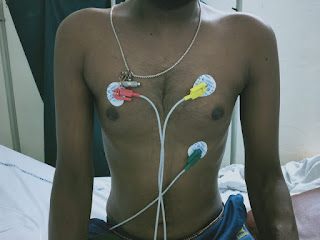
5.ECG


-irregular rhythm, absent p waves, right axis deviation,
-ST elevation in V4 V5 aVR


Cardiomegaly, enlargement of rt atrium, rt ventricle, Lt ventricle


-calcified mitral valves

-fish mouth appearance
based on the above investigations, the provisional diagnosis is MITRAL STENOSIS with HEART FAILURE.
TREATMENT:
1.INJ.LASIX 2amp in 50ml NS @8mg/hr
2.oxygenation to maintain spO2 above 95%
3.nebulization with budecort 12th hourly
4.strict I/O charting
5.monitoring BP,PR hourly
6.fluid and salt restriction
7.head end elevation
8.inj.amiodarone 300mg (2amp) at 6ml/hr
9.inj.pantop 40mg/OD/iv
10.T. ecosprin 75mg/PO/OD
(some insight in the case's history= patient was a agricultural labourer 5 years ago, mostly into plantation in paddy fields, occasionally did labour of carrying the harvest, which eventually caused her weakness and she had to quit!)


Comments
Post a Comment